Muscular System Diagram
A clear, labeled diagram of the major muscles of the human body, front and back. Click any muscle for its location and main action, switch to quiz mode to hide the labels and test yourself, and print a labeled or blank worksheet.
- Free to use
- No sign-up
- Runs in your browser
Explore the muscular system
Click a muscle or a label to learn about it.
Muscular System Study Pack
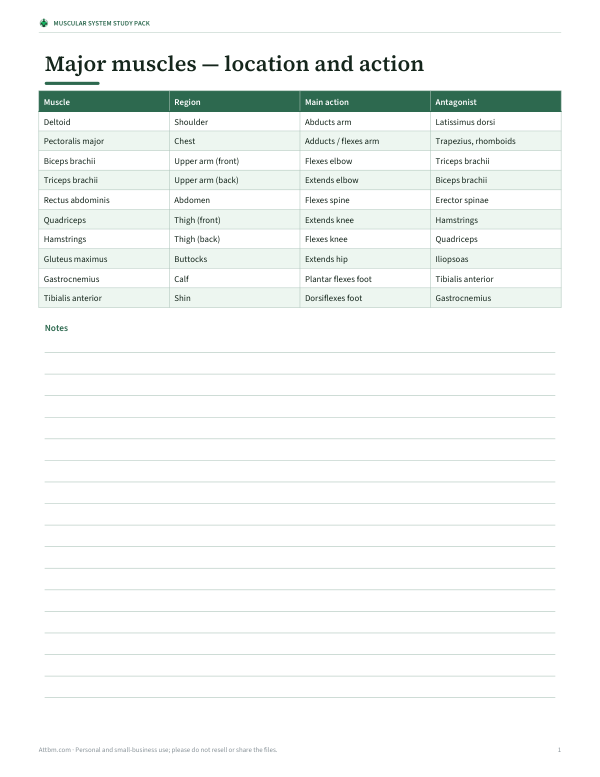
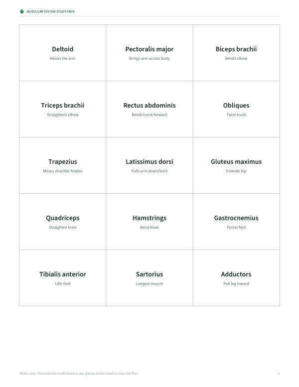
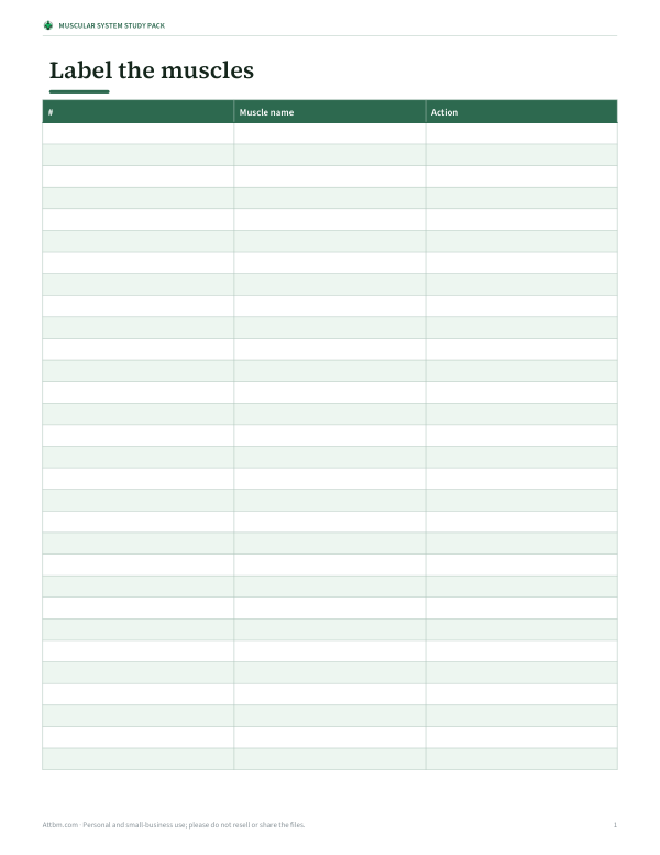
Printable labeled and blank muscular system worksheets, muscle flash cards with actions, and a muscle origin/action reference table.
- Muscle reference table (PDF, XLSX)
- Muscle flash cards (PDF, DOCX)
- Labeling worksheet (PDF, DOCX)
Formats: PDF, XLSX, DOCX. Instant download after payment (link valid 72 hours, up to 5 downloads). AI-assisted: the templates were drafted with AI help and reviewed and laid out before publishing.
$3.00 USD, one-time 3 files · about $1.00 each
- Instant download
- 7-day refund
- Secure checkout by Stripe
7-day money-back guarantee. Full refund within 7 days of purchase, even after you download.
Full refund within 7 days — see the refund policy and license.
Free printables
Single sheets you can print right now, no sign-up. The full pack adds every sheet in one download plus the editable files.
About this muscular system diagram
The human body has over 600 skeletal muscles. This diagram shows the major superficial muscles — the ones closest to the skin that you can see and feel — in a simplified front and back view, which is how they are usually taught in school biology, anatomy and physiology courses, and fitness qualifications. The shapes are schematic rather than photographic, so the focus stays on where each muscle is and what it does. Click a muscle to read its location and main action, or tick quiz mode to hide the names and test yourself.
Major muscles of the front of the body
| Muscle | Location | Main action |
|---|---|---|
| Deltoid | Shoulder | Raises the arm |
| Pectoralis major | Chest | Brings the arm across the body |
| Biceps brachii | Front of upper arm | Bends the elbow; turns palm up |
| Rectus abdominis | Front of abdomen | Bends the trunk forward |
| External obliques | Sides of abdomen | Twist and side-bend the trunk |
| Quadriceps | Front of thigh | Straighten the knee |
| Sartorius | Diagonal across thigh | Flexes and rotates hip; flexes knee |
| Adductors | Inner thigh | Pull the leg inward |
| Tibialis anterior | Front of shin | Lifts the foot |
Major muscles of the back of the body
| Muscle | Location | Main action |
|---|---|---|
| Trapezius | Upper back and neck | Moves and stabilises shoulder blades |
| Latissimus dorsi | Mid and lower back | Pulls the arm down and back |
| Triceps brachii | Back of upper arm | Straightens the elbow |
| Gluteus maximus | Buttocks | Extends the hip |
| Hamstrings | Back of thigh | Bend the knee; extend the hip |
| Gastrocnemius | Calf | Points the foot |
| Achilles tendon | Back of ankle | Attaches calf to heel |
How muscles work in pairs
Skeletal muscles can only pull, not push, so they are arranged in opposing pairs around joints. When one muscle — the agonist — contracts to move a joint, the opposite muscle — the antagonist — relaxes. The biceps and triceps are the classic example: the biceps bends the elbow, the triceps straightens it. The quadriceps and hamstrings do the same for the knee, and the tibialis anterior and calf muscles for the ankle. Understanding pairs helps with learning names and with planning balanced exercise.
Three types of muscle tissue
- Skeletal muscle — attached to bones by tendons; voluntary; striped (striated) under the microscope. These are the muscles on the diagram.
- Cardiac muscle — found only in the heart; involuntary; striated with branching cells.
- Smooth muscle — in the walls of organs and blood vessels, such as the stomach, intestines and arteries; involuntary; not striated.
Tips for learning muscle names
- Learn by region: shoulder and arm, chest and abdomen, back, hip and thigh, lower leg.
- Use the Latin clues: “brachii” means of the arm, “femoris” of the thigh, “maximus” largest, “rectus” straight, “oblique” slanted.
- Point to the muscle on your own body and contract it while saying the name.
- Use quiz mode and the printed blank worksheet for active recall.
- Learn each muscle with its action: “biceps — bends the elbow”.
Worked example: which muscles work in a squat?
Standing up from a squat, the quadriceps straighten the knees and the gluteus maximus extends the hips, with the hamstrings helping at the hip. The calf muscles and tibialis anterior stabilise the ankles, and the abdominals and back muscles keep the trunk upright. Mapping a familiar movement to the diagram is a good way to remember both the location and the action of each muscle.
Tendons, ligaments and how muscles attach
Muscles attach to bones through tendons, tough cords of connective tissue; ligaments, by contrast, connect bone to bone and stabilise joints. Each muscle is usually described by its origin — the more fixed attachment — and its insertion — the attachment that moves when the muscle contracts. The biceps brachii, for example, originates at the shoulder blade and inserts on the radius of the forearm, which is why contracting it bends the elbow and turns the palm upward. Learning origin and insertion is the next step after learning names and actions, and it explains why muscles produce the movements they do.
Muscles and exercise
Strength training works muscles against resistance so they adapt and grow stronger, while endurance activity improves their ability to work for long periods. Balanced programmes train opposing groups — chest and back, quadriceps and hamstrings — to support posture and reduce injury risk. Warming up increases blood flow and prepares muscles and tendons, and gradual progression allows tendons to adapt as well as muscles. For health-related advice about exercise or injury, speak to a qualified professional.
Using the printable worksheet
Print the labeled version as a study sheet or poster, and the quiz version — with numbered blank lines — as a worksheet or test. Teachers can project the diagram and switch between views while students label their own copies. The diagram is simplified for learning; for clinical or detailed anatomical study, use a full anatomy atlas.
Frequently asked questions
How many muscles are in the human body?
Over 600 skeletal muscles, depending on how they are counted.
What is the largest muscle?
The gluteus maximus is generally considered the largest by mass.
What is the longest muscle?
The sartorius, which runs diagonally across the front of the thigh.
Why do some charts label muscles differently?
Some charts group muscles (for example “quadriceps” for four muscles) while others show each one; anatomical names are the same, but the level of detail varies.
What is the strongest muscle?
It depends on how strength is defined; the masseter (jaw) produces very high force for its size, while the gluteus maximus and quadriceps generate the most total force.
What are antagonistic muscle pairs?
Muscles that work in opposition, such as the biceps and triceps.
Is this diagram accurate enough for school?
Yes for identifying major superficial muscles and their actions; it is simplified and schematic.
Can I print a blank version?
Yes, tick quiz mode and print for a numbered worksheet.